Be amazed at your eyes! They can show you extraordinary views, daily events, and the people you adore. The eyes are one of our body’s most intricate and essential parts. Let’s discover how every part works together to give us vision! You’ll find out why eyes are so miraculous!
Your eyes are a complex and marvelous part of your body. The size, shape, and structure let you see the world vividly. You must know how your eye works to appreciate your vision and maintain your visual health. In this guide, we’ll look at the anatomy of the eye—from its cornea to its lens.
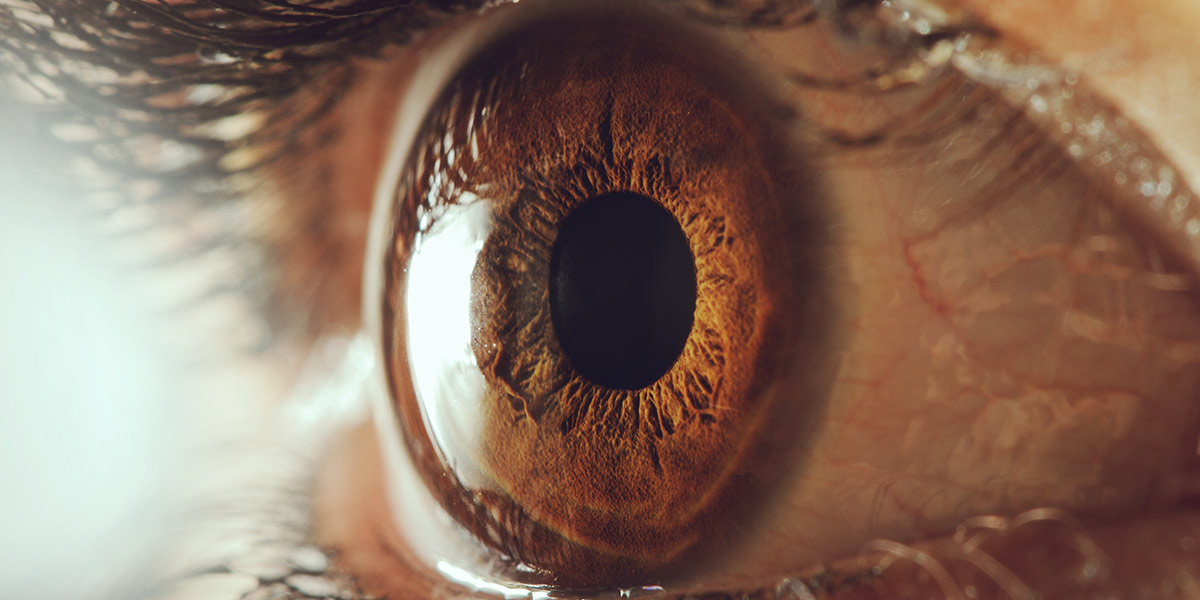
Your eye sits inside the skull’s orbital socket, which protects the delicate structures within. In your eye are several parts that work together to give you sight:
- Cornea: A transparent dome-shaped layer at the front of the eye that helps focus light waves.
- Iris: A colored, circular membrane inside the cornea with an opening (the pupil) that let’s light into the eye. It responds to changes in light intensity by contracting its muscles.
- Pupil: A dark hole in the center of the iris that allows light into the eye. It widens in dim conditions and shrinks in bright lighting.
- Lens: A clear disc behind the pupil that helps direct and focuses light waves onto the back of the eyeball.
- Vitreous humor: A transparent jelly-like material between the lens and retina. It keeps the eyeball shape by balancing pressure.
- Retina: A thin, multi-layered surface at the back of the eyeball. It has millions of photoreceptors (rods and cones) that convert light into electrical signals sent to the brain via the optic nerve.
Anatomy of the Eye
The eye is a complex organ made up of several parts. It begins with the cornea, and this tissue layer, shaped like a dome, allows light to enter. The iris follows, giving eyes their color and changing the size of the pupil to manage light.
The lens is next. It bends light rays for the retina to convert into an electrical signal. This signal is sent to the brain via the optic nerve so you can see. Tears come from the lacrimal glands to keep your eyes moist and washed.
Extraocular muscles control all eye motion. They attach to each side of the eyeballs and areas in your head and neck. You could look up, down, left, or right with them, and you’d be stuck looking straight ahead!
The Cornea
The cornea is a curved, transparent window on the front of the eye, and it helps light enter and helps focus on nearby objects. Its five main parts include:
- The epithelium is a thin layer of cells between the tear film and Bowman’s membrane. Bowman’s membrane protects cells from damaging elements like UV rays.
- The stroma is 90% of the cornea’s thickness and provides strength.
- Descemet’s membrane keeps fluids out while letting in vitamins, minerals, and other materials.
- Lastly, special Endothelium cells pump out excess fluid, preserving the cornea’s transparency.
The Iris
The iris is a prominent part of the eye. It’s found in the front, and its color and pattern give us our eye color. The iris controls how much light enters the pupil, and pigment cells inside the iris help regulate this. The pupil’s size changes depending on light levels and near or far-sightedness. The pupil’s size affects how much light comes in and how well we can see in the sun or dim rooms.
The Pupil
The pupil is the black, circular opening in the center of the eye. It acts as an adjustable aperture to regulate how much light enters and changes size depending on how bright it is outside. It dilates and constricts to maintain vision in different levels of light.
The iris surrounds the pupil. It’s a ring-like muscle that regulates the size and adds color to our vision. The iris has fibers that come together in a radial pattern. They are connected to muscles that attach to the ciliary body. These fibers help contract and expand. This decreases or increases the size of the pupil. It depends on the ambient light conditions.
The Lens
The lens is a biconvex, transparent structure in the eye. It lies behind the iris and pupil and is attached to muscles. It is made of proteins. Fibers attach to circles within the lens. This helps it keep its shape and change focus. Age can cause decreased stress, which is called presbyopia or farsightedness.
Cataracts, a clouding of lens tissue, can also occur due to age. Conditions like diabetic retinopathy or high blood pressure can damage the lens, leading to vision loss. New advances like an intraocular lens (IOL) implants can help replace damaged lenses with artificial replacements. This allows people to maintain normal vision after cataract surgery.
The Retina
The retina is a thin layer of tissue in the back of the eye. It is made up of several layers. These layers contain cells that sense light and turn it into electric signals. These signals travel through the retina’s nerve fibers, via the optic nerve, to your brain. Your brain interprets these signals as images.
The cells in the retina are known as rods and cones. Rods sense dim light, while cones sense bright light. They also control color vision. There is an oval-shaped pigmented area in the retina near its center. This area is called the macula. It contains many cones, giving a sharp central vision for fine details.
The retina also includes other structures, such as:
- Retinal pigment epithelium (RPE) cells nourish and maintain the photoreceptor cells.
- Ganglion cells send electrical signals along the optic nerve.
- Amacrine cells process visual information.
- Bipolar cells pass messages from photoreceptors to ganglion cells.
All these layers allow the eye to take in the world. You can enjoy moments without trying too hard!